Saving the Brain After a Stroke
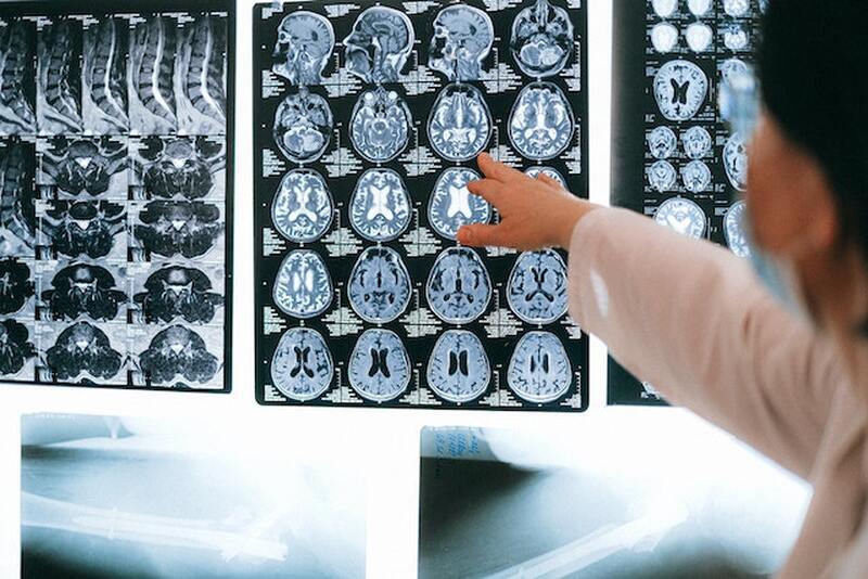
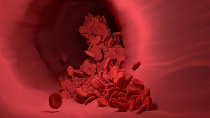
With one person dying from stroke every 3.5 minutes in the United States, strokes are one of the leading causes of death. While not all strokes are lethal, the rate of recurrence is high, with one in four stroke victims having previously had a stroke. A stroke occurs when a part of the brain suddenly does not receive enough crucial oxygen and nutrients carried by the blood, causing cells to die within minutes. The lack of nutrients to the brain can be a result of a hemorrhage, or rupture, in a blood vessel that supplies the brain, thus reducing the flow of blood. However, approximately 87% of strokes are ischemic strokes, which are due to a blood clot blocking a blood vessel that supplies parts of the brain, preventing cells from receiving the oxygen and nutrients that they need. Symptoms of a stroke often take place suddenly and include numbness of the limbs, confusion, and lack of coordination. These symptoms can vary from case to case and are dependent on the location of the stroke within the brain.
Because parts of the brain die from decreased blood flow, strokes can result in long-term disability. These damaged regions of the brain are called lesions. In an ischemic stroke, the lesion is composed of an ischemic core, where there is a dramatic decrease in blood flow, and the ischemic penumbra, a region surrounding the core that is at risk of damage but still salvageable and fully functional if properly treated. If doctors can save the penumbra in stroke patients, they can minimize impairments caused by the lesion. However, improper treatment or lack of treatment can lead to the penumbra becoming part of the core, the region of dead cells. Based on previous research, one of the main roadblocks in rescuing the penumbra is excitotoxic cell death, resulting from the neuronal release of an excessive amount of the neurotransmitter glutamate, which can damage different molecules within the cell. A recent study from Johannes Gutenberg University Mainz sought to find a new therapeutic method for saving the penumbra.
Because parts of the brain die from decreased blood flow, strokes can result in long-term disability. These damaged regions of the brain are called lesions. In an ischemic stroke, the lesion is composed of an ischemic core, where there is a dramatic decrease in blood flow, and the ischemic penumbra, a region surrounding the core that is at risk of damage but still salvageable and fully functional if properly treated. If doctors can save the penumbra in stroke patients, they can minimize impairments caused by the lesion. However, improper treatment or lack of treatment can lead to the penumbra becoming part of the core, the region of dead cells. Based on previous research, one of the main roadblocks in rescuing the penumbra is excitotoxic cell death, resulting from the neuronal release of an excessive amount of the neurotransmitter glutamate, which can damage different molecules within the cell. A recent study from Johannes Gutenberg University Mainz sought to find a new therapeutic method for saving the penumbra.
Image Source: Vector8DIY
The researchers examined autotaxin (ATX), an enzyme created by astrocytes, which are cells that surround synapses (gaps between neurons). They found that ATX is correlated with higher ischemic core volume, as ATX produces a molecule that promotes the release of glutamate and can contribute to excitotoxic cell death. Additionally, ATX was shown to be found at higher levels in both the brains of ischemic stroke mouse models and samples from human stroke victims. To see if blocking ATX could rescue the tissue within the penumbra, mice were given daily injections of an ATX inhibitor following a stroke. The researchers found that the inhibitor could significantly decrease the core volume and increase the amount of the penumbra saved. Furthermore, the inhibitor eventually improved the neurological function of the mice compared to untreated control mice, even when given up to 180 minutes after stroke. This result is significant because strokes should be treated as soon as possible to minimize brain damage, and despite the long 180-minute delay before treatment, the ATX inhibitor still improved neurological function in these mice.
The results from this study demonstrate a possible avenue for better treatment of stroke. Current standards of treatment for ischemic stroke involve directly removing the blood clot; however, little is done to save the still-repairable penumbra. Although the study primarily focused on mouse models of stroke, the researchers hope that their findings can spur additional research into ATX inhibition as an option for better stroke treatment.
The results from this study demonstrate a possible avenue for better treatment of stroke. Current standards of treatment for ischemic stroke involve directly removing the blood clot; however, little is done to save the still-repairable penumbra. Although the study primarily focused on mouse models of stroke, the researchers hope that their findings can spur additional research into ATX inhibition as an option for better stroke treatment.
Featured Image Source: Anna Shvets
RELATED ARTICLES
|
Vertical Divider
|
Vertical Divider
|
Vertical Divider
|


